The laboratory of Igor Koralnik, MD, in the Department of Neurological Sciences studies how the polyomavirus JC (JC virus) causes progressive multifocal leukoencephalopathy (PML). We study the clinical, radiological, immunological, histological and molecular aspects of this disease.
Our work
The virus: JC virus
- Non-enveloped, double-stranded DNA virus, papovaridae family
- Primary infection occurs during childhood, probable urine-oral route, up to 80 percent population is sero-positive
- Quiescent infection of kidney epithelium and lymphoid tissues
The disease: PML
- JCV reactivation in immunocompromised/HIV patients
- Up to 5 percent of individuals with AIDS prior to cART
- Patients treated with monoclonal antibodies for lymphoma (Rituximab/Rituxan), multiple sclerosis or Crohn's disease (Natalizumab/Tysabri), patients with hematological malignancies, or having undergone bone marrow or solid organ transplants
- Lytic infection of oligodendrocytes, resulting in demyelination and associated neurological deficits
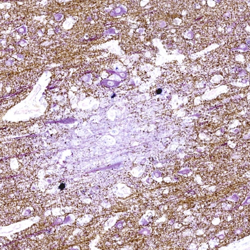
Presence of JCV-infected oligodendrocytes (blue) in a PML lesion at the gray/white junction. As JCV-infected oligodendrocytes are destroyed by the virus, their myelin sheaths (brown) wrapped around neurons (purple) disappear. Thin, purple neuronal processes, no longer protected by the myelin sheaths are visible in the lesion. Due to lack of myelin, the neuronal influxes in the PML lesions are greatly impaired.
The disease: PML-IRIS
Although the disease was originally considered to be devoid of inflammation, since the advent of combined antiretroviral therapy, or cART, we are confronted with PML in the setting of an immune reconstitution inflammatory syndrome, or IRIS. PML-IRIS is a paradoxical worsening or occurrence of new symptoms when the immune system is recovering. In the setting of HIV disease, this is often accompanied by a rise in CD4 count and a decrease in HIV viral load.
All natalizumab-associated PML cases have an IRIS phenomenon. Cytotoxic T-cell infiltrates are found in brains of PML-IRIS patients.
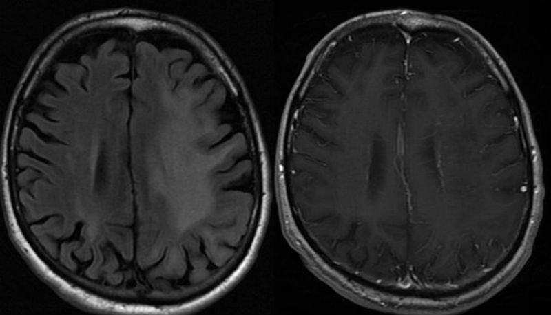
This is an MRI of the brain of a PML survivor. On the left is a fluid attenuated inverse recovery, or FLAIR, image, which shows a large hyperintense lesion in the left cerebral hemisphere, sparing the cortex. On the right is the corresponding T1 with gadolinium (contrast) image, which shows no enhancement within the hypointense PML lesion.
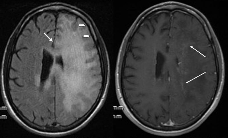
This is an MRI of the brain of a PML-IRIS survivor. On the left, FLAIR image shows a large hyperintense lesion in the white matter of the left hemisphere. There is also a shift of the midline (arrow) and disappearance of the sulci (arrowheads), signifying mass effect, as seen in excessive inflammation. On the right is the corresponding T1+Gad image; the arrows show enhancement within the PML lesion.
Discovery of three novel diseases associated with JC virus
In addition to PML, the Koralnik laboratory has characterized novel clinical syndromes caused by JC virus infection of neurons and meningeal cells: JCV granule cell neuronopathy (JCV GCN), JCV encephalopathy (JCVE) and JCV meningitis (JCVM).